
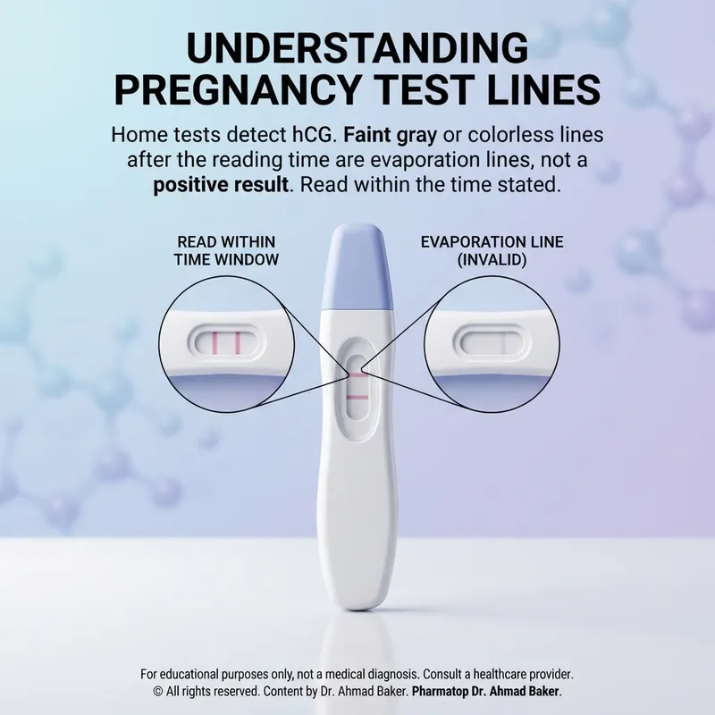
Analytical Chemistry of Lateral Flow hCG Immunoassays: Resolving Evaporation Artifacts, Kinetic Windows, and Diagnostic Ambiguity
Clinical Abstract: Point-of-care (POC) and home urine pregnancy tests rely on qualitative chromatographic lateral flow immunoassays (LFIA) designed to capture intact human Chorionic Gonadotropin (hCG). A persistent source of diagnostic confusion is the delayed appearance of an indeterminate, colorless, or grayish band after the validated reading window has elapsed—physicochemically identified as an evaporation line. This clinical monograph examines the molecular biology of hCG isoforms, the physical chemistry of nitrocellulose membrane desiccation, chromatographic capillary flow kinetics, hook effects, cross-reactivity profiles, and evidence-based clinical verification algorithms.
📑 Comprehensive Monograph Contents
⚡ Diagnostic Immunoassay Factsheet
| Target Biomarker | Human Chorionic Gonadotropin (Intact Dimeric hCG, β-subunit) |
| Assay Methodology | Qualitative Sandwich Lateral Flow Chromatographic Immunoassay |
| Standard Analytical Sensitivity | 10 mIU/mL to 25 mIU/mL (calibrated against WHO International Standard) |
| Validated Reading Window | Strictly 3.0 to 5.0 minutes (Maximum allowable: 10.0 minutes) |
| Optical Label Technology | Colloidal Gold Nanoparticles (Pink/Red) or Dyed Polystyrene Latex (Blue) |
| Renal Biomarker Half-Life | Circulating intact hCG t½ ~24–36 hours; rapid urinary excretion of metabolites |
1. Molecular Biology & Heterogeneity of Human Chorionic Gonadotropin (hCG)
Human Chorionic Gonadotropin (hCG) is a heterodimeric glycoprotein hormone synthesized predominantly by syncytiotrophoblast cells following blastocyst implantation. The intact molecule possesses a molecular weight of approximately 36.7 kDa and comprises two non-covalently linked polypeptide subunits:
- Alpha (α) Subunit: Comprising 92 amino acids, this subunit is structurally identical to the α-subunits of other anterior pituitary glycoprotein hormones, specifically Luteinizing Hormone (LH), Follicle-Stimulating Hormone (FSH), and Thyroid-Stimulating Hormone (TSH).
- Beta (β) Subunit: Comprising 145 amino acids, the β-subunit confers unique biological and immunological specificity. It contains a distinctive 32-amino acid carboxyl-terminal peptide (CTP) with four O-linked oligosaccharide chains, preventing non-specific cross-reactivity in modern diagnostic assays directed against specific β-epitopes.
In biological fluids, hCG exhibits extreme molecular microheterogeneity. Rather than circulating solely as an intact heterodimer, several metabolic variants coexist in serum and urine:
- Hyperglycosylated hCG (hCG-h): Synthesized by extravillous cytotrophoblasts; represents the dominant isoform in early post-implantation gestation (weeks 3 to 5).
- Nicked hCG (hCGn): Formed via peptide bond cleavage within the β-subunit core loop (typically residues β44–45 or β47–48) by macrophage proteases.
- Free Beta Subunit (free β-hCG): Uncomplexed monomeric β-chains, prominent in early embryogenesis and certain trophoblastic neoplasms.
- Beta-Core Fragment (hCGβcf): A low-molecular-weight degradation product (~10 kDa) consisting of disulfide-linked segments of the β-subunit (residues 6–40 linked to 55–92). It represents the predominant immunoreactive urinary species after the 6th week of gestation.
Commercial qualitative assays must demonstrate balanced affinity toward intact hCG and hyperglycosylated hCG while avoiding false-negative saturation from excessive concentrations of urinary hCGβcf.
2. Lateral Flow Immunoassay (LFA) Chromatography & Reaction Kinetics
The qualitative home and point-of-care pregnancy test is built upon a membrane-based lateral flow chromatographic immunoassay format, utilizing a standard “sandwich” architecture across four discrete zones:
- Sample Application Pad: A porous woven mesh (cellulose or glass fiber) that buffers the sample, filters cellular debris, and maintains an optimal pH (typically 7.2–7.6) for antibody-antigen binding.
- Conjugate Release Pad: Impregnated with mobile, highly specific monoclonal anti-hCG antibodies (typically murine IgG targeting an epitope on the β-subunit). These antibodies are covalently coupled to optical reporter particles—most commonly colloidal gold nanoparticles (producing a crimson/pink spectral band via surface plasmon resonance) or dyed polystyrene latex microspheres (producing a distinct blue band).
- Nitrocellulose Analytical Membrane: A capillary-active porous polymer matrix with precisely defined pore sizes (5–15 µm) that houses two immobilized reagent lines:
- Test Zone (“T” Line): Consists of fixed, irreversibly bound anti-hCG capture antibodies (monoclonal or polyclonal, targeting an independent epitope on the α-subunit or a non-overlapping β-domain).
- Control Zone (“C” Line): Consists of immobilized secondary species-specific antibodies (e.g., goat anti-mouse IgG) that capture excess mobile conjugate particles regardless of analyte presence, validating capillary fluid migration.
- Absorption (Wick) Pad: A high-capacity cellulosic reservoir situated at the distal end that drives capillary draw and prevents fluid backflow (retrograde migration).
Chromatographic Mechanism:
Sample Hydration → Solubilization of Gold-Anti-β-hCG Conjugate → Formation of [hCG:Gold-Ab] Complex → Capillary Migration across Nitrocellulose → Capture at Test Line by Anti-α-hCG → Formation of Sandwich Complex [Ab_capture : hCG : Ab_conjugate-Gold] → Visible Chromatic Band
3. The Biophysics of Evaporation Lines vs. True Immunoassay Bands
A frequent challenge in point-of-care diagnostics is the clinical differentiation between a trace-positive immunoassay result and a post-reaction physical artifact, termed an evaporation line.
A true positive reaction represents a specific biochemical event. The optical reporter nanoparticles (colloidal gold or colored latex) are immobilized at the Test Zone via high-affinity immunological binding (Kd ~ 10⁻⁹ to 10⁻¹¹ M). This creates a distinct, continuous band possessing the specific dye hue (pink/red for gold, blue for latex).
Conversely, an evaporation line is a non-immunological artifact that develops as the nitrocellulose matrix dries. The biophysical mechanism involves several phenomena:
- Capillary Desiccation & Meniscus Collapse: As water evaporates from the porous nitrocellulose strip over 30 to 120 minutes, the liquid meniscus recedes, leaving the membrane dry.
- Solute Crystallization: Normal urinary constituents—including urea, uric acid, Tamm-Horsfall mucoproteins, and inorganic salts—precipitate directly onto the membrane as their solubility thresholds are exceeded.
- Optical Phase Boundary Distortion: The mechanical scoring or chemical pre-treatment at the Test Zone alters the local refractive index of the nitrocellulose. When ambient light strikes this line of dry protein-mineral precipitate, light scattering produces a faint, shadow-like, grayish, or colorless band that mimics a positive result.
- Retrograde Capillary Flow: As the wick pad reaches equilibrium and the strip begins to desiccate, unreacted, unbound conjugate nanoparticles may slowly migrate in reverse, settling non-specifically into the physical depression of the Test line.
| Diagnostic Parameter | True Positive Result | Evaporation Line (Artifact) |
|---|---|---|
| Appearance Time | Within validated window (3–5 min) | Delayed (>15–30 min to hours) |
| Chromatic Integrity | Clear pink, red, or blue pigment | Colorless, gray, yellowish, or shadow-like |
| Morphological Width | Uniform, complete, matches Control line | Thin (“hairline”), uneven, or partial edge |
| Hydration Status | Appears while membrane is wet | Appears only as membrane becomes desiccated |
| Clinical Significance | Presence of β-hCG ≥ assay cut-off | Diagnostic invalidity (Negative or Indeterminate) |
💬 Require Professional Guidance on This Medication?
Dr. Ahmad Baker and the clinical advisory team at Pharma Guide provide evidence-based, confidential pharmacological consultations.
Request Consultation4. Diagnostic Kinetic Windows & Optical Resolution Timelines
The kinetic performance of a lateral flow device is governed by the principles of capillary action and antibody-antigen association rates ($k_{on}$) versus dissociation rates ($k_{off}$). The testing timeline can be categorized into three distinct phases:
- Phase 1: Capillary Inflow & Solubilization (0 to 120 seconds): The sample hydrates the conjugate pad, mobilizing antibody-gold nanoparticles. The fluid moves along the strip, passing the Test and Control lines. The membrane appears transiently pinkish or wet, reflecting light as the background clears.
- Phase 2: Thermodynamic Equilibrium & Optimal Read Window (3 to 5 minutes): The background clears to a crisp, uniform white. Bound analyte-antibody complexes reach peak optical density. This represents the only validated window for accurate diagnostic interpretation.
- Phase 3: Desiccation & Non-Specific Aggregation (10 minutes to several hours): Capillary pull ceases as the absorbent wick saturates. Ambient evaporation drives local solute deposition. Any band that develops during this phase must be classified as an unvalidated artifact.
Modern digital immunoassay readers mitigate human interpretation error by incorporating optoelectronic reflectance sensors. These units measure light reflectance at specific wavelengths precisely at the 3-minute mark, translating optical density into an unambiguous alphanumeric readout (e.g., “Pregnant” vs. “Not Pregnant”) and eliminating subjective evaluations of faint lines.
5. Biological & Assay Interferences: False Positives and Negatives
Understanding biological and analytical interferences is essential for correctly interpreting ambiguous or anomalous results:
Etiologies of True Analytical False Positives:
- Heterophile Antibodies & Human Anti-Mouse Antibodies (HAMA): Non-specific human immunoglobulins that cross-link the murine-derived capture and conjugate antibodies in the absence of hCG, triggering false-positive band formation.
- Pituitary hCG Secretion: In perimenopausal and postmenopausal women, declining ovarian estrogen feedback stimulates pituitary gonadotropes to secrete small amounts of pituitary hCG (typically 5–15 mIU/mL) alongside elevated LH levels. Highly sensitive assays (10 mIU/mL cutoff) may register these low baseline levels.
- Gestational Trophoblastic Disease (GTD): Hydatidiform moles, invasive moles, and choriocarcinomas secrete massive quantities of heterogeneous hCG variants.
- Non-Trophoblastic Neoplasms: Ectopic production of β-hCG by certain malignancies (e.g., germ cell tumors, ovarian, gastric, or neuroendocrine cancers).
- Early Pregnancy Loss (Chemical Pregnancy): Detection of residual β-hCG following very early embryonic loss occurring shortly after implantation.
Etiologies of True Analytical False Negatives:
- The High-Dose Hook Effect (Prozone Phenomenon): Occurs when extremely high concentrations of intact hCG (typically >500,000 mIU/mL, as seen in advanced molar pregnancies or multiple gestations) saturate both the mobile conjugate and fixed capture antibodies independently, preventing the formation of the visual sandwich complex and causing a falsely faint or negative Test line.
- The Variant Hook Effect (hCGβcf Interference): High concentrations of urinary hCG beta-core fragments (which lack the intact α-subunit) bind to capture or conjugate antibodies without completing the sandwich complex, suppressing signal generation in mid-to-late first-trimester samples.
- Premature Testing: Testing prior to the doubling of maternal serum and urinary concentrations above the assay’s lower limit of detection (LLOD).
🚨 Urgent Clinical Alert: Immediate Emergency Criteria
Immediate quantitative serum testing and emergency gynecological evaluation are mandatory if an ambiguous or positive pregnancy test is accompanied by any of the following symptoms:
- Severe Lateralized Pelvic or Abdominal Pain: Cardinal presentation suggestive of an unruptured or ruptured tubal ectopic pregnancy.
- Hemodynamic Instability: Syncope, severe orthostatic dizziness, hypotension, or unexplained tachycardia.
- Referred Shoulder Tip Pain: Diaphragmatic irritation secondary to hemoperitoneum (intra-abdominal bleeding).
- Abnormal Uterine Hemorrhage: Significant vaginal bleeding accompanied by expulsion of tissue fragments or decidual casts.
6. Pharmacological & Exogenous Drug Interferences
Misinterpretation often stems from a lack of clarity regarding which pharmacological agents can biologically or chemically alter qualitative pregnancy test results:
- Exogenous hCG Administration: Patients undergoing Assisted Reproductive Technology (ART) or ovulation induction regimens receiving exogenous hCG (e.g., Novarel, Pregnyl, Ovidrel) will clear the hormone with a terminal half-life of 24 to 36 hours. Residual circulating hormone can trigger false-positive tests for up to 10–14 days post-injection.
- Biotin (Vitamin B7) Interference: High-dose dietary biotin supplementation (>5–10 mg/day) can interfere with laboratory and point-of-care immunoassays utilizing streptavidin-biotin capture architectures, potentially causing false-negative or falsely suppressed results.
- Non-Interfering Pharmaceuticals: Standard medications—including oral contraceptives, antibiotics (e.g., amoxicillin, azithromycin), analgesics (e.g., ibuprofen, acetaminophen), antipyretics, and antihistamines—do not cross-react with β-hCG epitopes and do not cause false-positive results or evaporation lines.
For detailed pharmacological monographs on reproductive medications, explore the Comprehensive Drug Index.
7. Matrix Effects, Hydration & Renal Clearance
Urinary matrix properties significantly influence lateral flow performance:
Urinary Specific Gravity & Osmolality: Ingesting large volumes of fluid prior to testing dilutes the urine, lowering specific gravity (<1.010) and reducing the concentration of intact hCG below the assay’s lower limit of detection (e.g., diluted to 8 mIU/mL when the threshold is 20 mIU/mL). This creates a false-negative result early in gestation. Testing with a first-morning urine sample provides the highest concentration of overnight analyte accumulation.
Proteinuria and Hematuria: Elevated levels of serum proteins (albuminuria >500 mg/dL) or red blood cells (gross hematuria) increase sample viscosity, slow capillary flow, and can cause non-specific binding along the nitrocellulose membrane, potentially creating faint non-specific background staining.
8. Clinical Verification: Quantitative Serum β-hCG & TVUS Correlation
When qualitative urine testing yields indeterminate results, clinical protocols transition to quantitative laboratory and imaging modalities:
Serial Quantitative Serum Total β-hCG: Automated chemiluminescent immunoassays (CLIA) quantify intact and subunit β-hCG down to <1–2 mIU/mL. In a viable early intrauterine pregnancy, maternal serum β-hCG levels typically rise predictably, with an expected doubling time of 48 to 72 hours (or a minimum rise of 35%–53% over 48 hours in very early gestations). An abnormal rise (plateauing) or an inappropriate decline suggests a non-viable pregnancy (spontaneous abortion) or an ectopic gestation.
The Transvaginal Ultrasonography (TVUS) Discriminatory Zone: The discriminatory zone represents the serum quantitative β-hCG threshold above which an intrauterine gestational sac should reliably be visualized via high-resolution TVUS. International consensus guidelines (ACOG/SMFM) establish this threshold between 1,500 and 2,000 mIU/mL (and up to 3,500 mIU/mL to prevent premature intervention). If serum levels exceed this zone without an identifiable intrauterine sac, providers must suspect an ectopic pregnancy or complete spontaneous miscarriage.
For broader clinical protocols on reproductive health diagnostics, visit the Women’s Specialized Clinical Care and Sexual & Reproductive Health Hub.
9. Manufacturing Quality, Shelf-Life & Membrane Degradation
The integrity of point-of-care immunoassay devices depends heavily on storage chemistry and manufacturing quality control:
- Nitrocellulose Aging: Over time, nitrocellulose undergoes progressive polymer degradation, altering pore uniformity, capillary migration rates, and background clarity.
- Conjugate Antibody Denaturation: Exposure to temperatures above 30°C (86°F) or elevated humidity damages the tertiary protein structure of the immobilized and conjugated antibodies, leading to non-specific aggregation or loss of binding capacity.
- Desiccant Pouch Failures: Defective foil packaging allows ambient moisture into the cassette, hydrating the conjugate pad prematurely and precipitating gold-antibody conjugates before sample application.
10. Clinical Pharmacist Assessment & Decision-Tree
When evaluating ambiguous pregnancy test results, clinical pharmacists should guide patients through a structured decision algorithm:
- Assess the Time Window: Did the line appear within 3 to 5 minutes? If the line developed after >10–15 minutes, document the result as unvalidated/indeterminate (likely an evaporation artifact).
- Evaluate Visual Characteristics: Does the band display clear, distinct color (pink/red/blue), or is it an irregular, grayish shadow? Only pigmented lines are considered valid positives.
- Protocol for Indeterminate or Evaporative Lines:
- Advise the patient to re-test after a 48-hour interval using a fresh, first-morning urine sample to allow endogenous β-hCG concentrations to double if early conception has occurred.
- Recommend an optoelectronic digital pregnancy test if visual interpretation remains ambiguous.
- Protocol for Persistent Ambiguity: Refer the patient for laboratory-based serial quantitative serum total β-hCG testing and clinical evaluation by a healthcare provider.
❓ Frequently Asked Clinical Questions
❓ Why do heterophile antibodies cause false-positive immunoassay results, and how is this distinguished from gestational hCG?
Heterophile antibodies (e.g., Human Anti-Mouse Antibodies, or HAMA) are endogenous immunoglobulins with non-specific binding affinity for animal-derived diagnostic antibodies. In sandwich immunoassays, they bind simultaneously to the murine conjugate and capture antibodies, forming an antibody-heterophile-antibody bridge without any hCG present. This artifact can be distinguished from true gestational hCG by serial serum dilution (which displays non-linear kinetics), urine qualitative testing (heterophile antibodies are rarely filtered across the glomerulus due to their high molecular weight), or pre-incubating the sample with heterophile-blocking tubes.
❓ What is the high-dose “hook effect” (prozone phenomenon) in pregnancy diagnostics?
The hook effect occurs when extremely high analyte concentrations (>500,000 mIU/mL, as seen in complete molar pregnancies or choriocarcinomas) overwhelm the immunoassay system. Excess uncomplexed hCG saturates both the mobile tracer antibodies and the stationary capture antibodies independently. This prevents the formation of the complete sandwich complex [Capture-Ab : hCG : Tracer-Ab], paradoxically yielding a falsely faint or entirely negative test line. This interference is resolved in the laboratory by performing serial sample dilutions.
❓ How does the “variant hook effect” driven by hCG beta-core fragments interfere with qualitative tests?
hCG beta-core fragment (hCGβcf) is the principal breakdown product of hCG in maternal urine during mid-to-late pregnancy. Because hCGβcf possesses the β-subunit epitope but lacks the α-subunit, it can bind to capture antibodies on the test line without completing the signal-generating sandwich. At high concentrations, it outcompetes intact dimeric hCG for binding sites, leading to falsely negative or faint results despite high total hCG levels.
❓ What biochemical distinction separates an evaporation artifact line from a faint true positive band?
A true positive band represents the immunochemical immobilization of optical reporter nanoparticles (colloidal gold or dyed latex), producing a distinct pink, red, or blue line across the entire width of the Test zone within the 3-to-5-minute read window. An evaporation line is a post-test physical artifact that occurs as the nitrocellulose dries (>15–30 minutes), caused by the precipitation of urinary salts and proteins that alter light refraction, producing a colorless, grayish, or shadow-like mark.
❓ How does exogenous hCG administered during ART protocols affect clearance kinetics?
Exogenous hCG (e.g., intramuscular or subcutaneous trigger injections of 5,000–10,000 IU) is eliminated following multi-exponential decay with a terminal elimination half-life of 24 to 36 hours. Depending on renal function, body mass index, and total dose, residual circulating concentrations can remain above the analytical sensitivity threshold of home pregnancy tests (10–25 mIU/mL) for up to 10 to 14 days post-administration, potentially causing false-positive readings.
❓ Can elevated pituitary hCG in perimenopausal women trigger false-positive tests?
Yes. With the decline of ovarian follicular function and loss of negative feedback from estradiol and progesterone, the anterior pituitary increases production of gonadotropins (LH and FSH), accompanied by low levels of pituitary-derived hCG (typically 3–15 mIU/mL). Highly sensitive qualitative assays with lower detection limits of 10 mIU/mL can detect this baseline secretion, producing a faint positive result in the absence of pregnancy.
❓ How does high-dose Biotin (Vitamin B7) supplementation interfere with modern pregnancy diagnostic platforms?
Many laboratory immunoassays utilize a biotin-streptavidin binding system to anchor capture antibodies to the solid phase. Ingesting high therapeutic or supplemental doses of Biotin (>5–10 mg/day) results in elevated free biotin concentrations in patient serum, which compete with biotinylated diagnostic antibodies for streptavidin binding sites. In sandwich immunoassays, this causes falsely low or false-negative results.
❓ What is the clinical significance of the “discriminatory zone” for serum β-hCG and TVUS?
The discriminatory zone is the serum quantitative total β-hCG concentration (established between 1,500 and 2,000 mIU/mL, and up to 3,500 mIU/mL in conservative guidelines) above which a normal, singleton intrauterine gestational sac must be reliably visualized via transvaginal ultrasonography. An absence of an intrauterine sac at or above this threshold raises high suspicion for an ectopic pregnancy, early pregnancy demise, or complete abortion.
❓ How does urinary specific gravity affect the analytical sensitivity of qualitative lateral flow assays?
High fluid intake dilutes the urine, reducing its specific gravity (<1.010) and lowering analyte concentration. In very early pregnancy, this dilution can drop urinary hCG levels below the assay’s detection limit (e.g., diluting a true level of 18 mIU/mL down to 6 mIU/mL against a 20 mIU/mL threshold), resulting in a false negative. Testing with a first-morning urine sample, which is naturally concentrated overnight, avoids this dilution effect.
❓ What is “phantom hCG,” and how is it clinically confirmed?
“Phantom hCG” refers to persistent, false-positive serum β-hCG test results caused by heterophile or human anti-animal antibodies in patients without a true pregnancy or trophoblastic disease. It is clinically confirmed when the patient demonstrates persistently elevated serum β-hCG levels that fail to appear in parallel urine samples (since large immunoglobulins cannot cross the glomerular filtration barrier) and fail to clear or drop following clinical interventions.
❓ How do anti-mouse antibodies (HAMA) interact with mouse-derived monoclonal diagnostic strips?
Human Anti-Mouse Antibodies (HAMA) develop following exposure to murine therapeutic antibodies or environmental antigens. Because lateral flow strips typically use murine monoclonal antibodies for both conjugate and capture phases, HAMA binds to the Fc domains of both antibodies, cross-linking them on the Test line. This generates a visible, colored band identical to a positive test result in the complete absence of hCG.
❓ Why does the alpha subunit of hCG share structural homology with LH, FSH, and TSH, and how is cross-reactivity prevented?
hCG, LH, FSH, and TSH evolved from a common ancestral gene and share an identical 92-amino acid α-subunit. Cross-reactivity in modern diagnostic assays is avoided by using monoclonal antibodies specifically directed against distinct structural epitopes on the unique 145-amino acid β-subunit—particularly the 32-amino acid carboxyl-terminal peptide (CTP)—ensuring no cross-reaction with circulating LH surges or elevated TSH.
📚 Clinical References & Authoritative Sources
- American College of Obstetricians and Gynecologists (ACOG). (2018). Tubal Ectopic Pregnancy. ACOG Practice Bulletin No. 193. Obstetrics & Gynecology, 131(3), e91-e103.
- Clinical and Laboratory Standards Institute (CLSI). (2021). Immunoassay Methods for the Assessment of Human Chorionic Gonadotropin (hCG); Approved Guideline. CLSI document I/LA30. Wayne, PA.
- U.S. Food and Drug Administration (FDA). Over-the-Counter Pregnancy Tests: Medical Device Guidance and Performance Criteria. Center for Devices and Radiological Health.
- Cole, L. A. (2012). hCG, the wonder of gestational hormones. Reference Module in Biomedical Sciences / Elsevier, 12(2), 115-127.
- Tietz Textbook of Clinical Chemistry and Molecular Diagnostics. (6th ed.). Human Chorionic Gonadotropin and Pregnancy Testing. St. Louis: Elsevier Saunders.
⚠️ Clinical Disclaimer
The clinical monographs published on Pharma Guide are intended strictly for medical education and research reference. They do not constitute individualized medical advice or diagnostic confirmation. Diagnostic ambiguity, suspected ectopic gestation, or discordant immunoassay findings must be evaluated under the supervision of a licensed physician or through clinical communication with Dr. Ahmad Baker. Review our complete Medical Disclaimer.
© 2026 Pharma Guide – Clinical Drug Index & Evidence-Based Diagnostic Protocols.
Under the Clinical Direction of Dr. Ahmad Baker | Editorial Policy | Medical Disclaimer | Contact Support

Dr. Ahmad Baker, PharmD
Senior Pharmacist & Health Educator
With over 23 years of clinical experience in hospital and community pharmacy across Egypt and Saudi Arabia, Dr. Ahmad specializes in reproductive health and pharmaceutical safety. He is dedicated to providing evidence-based guidance to empower patients in the MENA region.
Medical Disclaimer: This content is for educational purposes and reflects the latest pharmaceutical protocols. It is not a substitute for personal medical advice. Always consult Dr. Ahmad Baker or your healthcare provider before starting any treatment.